The Ultimate Guide to Moles
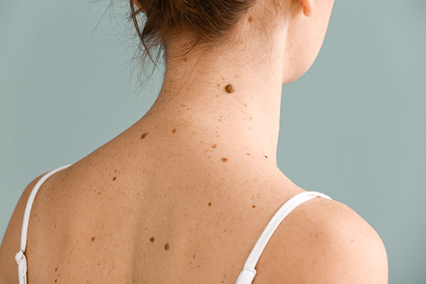
During childhood and adolescence, sun exposure affects the number and size of moles that may appear on the skin. The more moles emerge, the greater the likelihood of developing skin cancer, as 35% of skin cancers develop from pre-existing lesions. For this reason, it is crucial to effectively protect children and adolescents from the sun to reduce the number of moles as much as possible, since excessive exposure can also lead to cancerous changes. Hence, it is essential to develop good habits to protect moles from type A (UVA) and B (UVB) ultraviolet rays. UVA is linked to skin aging due to its longer wavelength, whereas UVB exposure is associated with skin burning.
Not all lesions are alarming, but even the harmless moles, especially if challenging to hide from the sun, are worth getting checked and possibly removed. The medical practitioner decides on the method of mole removal depending on the nature of the mole. On the basis of examining the changes – their color and shape, for instance – your physician can distinguish the “healthy” moles from those that can become troublesome or even cancerous.
Mission: Recognize Melanoma
One of the most characteristic melanoma symptoms is the appearance of a new mole or a change in color, shape, or size of a mole that we’ve had for a while. In fact, any change in the skin “deserves” consultation with a doctor.
A helpful diagnostic tool that we can use, even at home, is the ABCDE criteria.
Melanoma is a malignant tumor that arises from melanocytic (pigmented) cells. Most often, melanoma develops in the skin, but it can also appear in the eyeball and mucous membranes. To distinguish melanoma from a typical mole, the lesion should be evaluated for the following clinical features:
-
- Asymmetry – suspicious changes have asymmetrical dimensions. The healthy mole is rather symmetrical in shape;
- Border – lesions of a malignant nature have very irregular margins;
- Color – the healthy mole is usually uniform in color. In the case of melanoma-like lesions, the color is not consistent, has highlights or darkening on the lesion’s surface;
- Diameter – the abnormal size of the lesion, greater than 6mm;
- Evolving – noticeable changes in the lesion that occur rapidly, such as size, color, bleeding, thickness, or shape.
The primary method of diagnosing melanoma is dermatoscopy, a non-invasive and pain-free procedure consisting of inspecting the subsurface skin structures under magnification. These include the epidermis, the dermo-epidermal junction, and the upper dermis, all of which are usually not visible to the naked eye. The basis for further diagnosis is a biopsy, which is the microscopic examination of the lesion removed by a doctor.
Moles: to remove or not to remove?
“It’s better not to touch” is an archaic belief that may have serious consequences. The principle is quite the opposite – it is better to get rid of the irritating mole in advance. The harmful myth that some birthmarks or other skin lesions should not be touched, and that their excision may end tragically, was most likely caused by the patients visiting a specialist too late, when removing the mole could no longer help.
The excision of the whole lesion does not lead to neoplastic transformation and metastasis. Suppose metastases are found after a properly performed surgery. In that case, it may indicate that the removed lesion was a melanoma that had spread through the lymphatic and blood vessels before the surgery. Skin lesions are also often removed for aesthetic reasons, even if they turn out to be entirely harmless.
As we age, atypical moles continue to form and can be found on all parts of the body, especially in areas that are regularly covered. People who have many atypical moles and those with a family history of melanoma should be especially conscientious since the tendency is hereditary.
Important Note
The risk of developing melanoma includes people who have a family history of someone who suffered from the disease, frequent sunbathing, lack of sunscreen application, fair-skinned people, and those who suffered from sunburn in childhood. “Suspicious” moles should be examined by a doctor once a year; moreover, thorough self-examination should be performed every few months. If you don’t trust your memory, take pictures of the lesions – it will be easier to see if the moles have changed in any way.
Mole Removal: laser or surgery
There are several methods of mole excision. With many skin lesions, warts, and fibromas, removal can be performed using a laser. During the treatment, the laser energy is absorbed by the tissue where water evaporates, and coagulation occurs. This way, there is no bleeding. In addition, the laser does not cause thermal damage to the tissue around the lesion. The solutions used in the process positively affect the healing process that takes place without scarring.
The surgical method consists of cutting out the lesion with a scalpel with local anesthesia – ointment or injection. The moles are removed with a margin of healthy skin to ensure that no potentially dangerous tissue remains. After the mole is removed, sutures and dressing are applied.